


Stay up to date on upcoming events, deadlines, news, and more by signing up for our newsletters!
MRI scans are a cornerstone of modern medicine, but they're notoriously slow. This bottleneck can mean a longer, more anxious wait for patients needing diagnoses for strokes or brain tumors. Two NYU PhDs, Dr. Gregory Lemberskiy (Grossman '19) and Dr. Benjamin Aron (Tandon '22), decided to tackle this problem head-on. Their startup, MICSI, is deploying a revolutionary AI that can cut scan times in half and dramatically increase image resolution, redefining the boundaries of diagnostic imaging.
Why do MRIs work so slowly? A lot of time is spent on preserving an acceptable signal-to-noise ratio, a process that often involves acquiring an image one line at a time like a fax machine. MICSI’s AI technology, on the other hand, works via a “smart averaging” approach. It combines multiple MRI scan images, identifying both their unique image properties and their noise levels in order to enhance the former and discard the latter. MICSI-RMT is the first commercial software built on a powerful denoising algorithm pioneered by NYU Grossman Radiology professors Jelle Veraart, Els Fieremans, and Dmitry Novikov. Unlike other AI, it doesn't require massive external datasets or supercomputers to work. Using MICSI-RMT, MRI centers can produce higher resolution imaging and reduce scan times by as much as 50 percent. This means more patients scanned, earlier disease interventions, and lives saved.
The team’s aspirations don’t stop there. “Radiology is an art project,” says Aron. “All the images are anatomic. They give you a pretty picture of the brain and many companies are saying, how can we make that picture clearer? While we do improve the image resolution, our goal is to also improve the image's accuracy and precision - the sensitivity of that image to disease as a biomarker.” Higher image accuracy means deeper insight into changes in a patient’s cellular microstructure. “You can detect these changes much sooner than you can detect anatomy,” explains Lemberskiy. “Say, for example, cellular beading, like how the axons swell up prior to stroke. The holy grail is for MICSI to be able to produce imaging biomarkers that use microstructural information.”
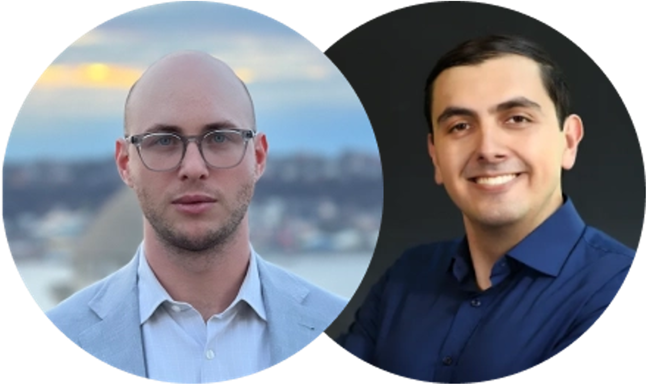
MICSI Founders Gregory Lemberskiy & Benjamin Aron
MICSI emerged from the work Aron and Lemberskiy collaborated on as researchers at the NYU Grossman School of Medicine. The initial driving impulse was sheer practicality. “As a scientist building something new, what do you do? Everyone says, well, you apply for a grant,” recalls Lemberskiy. “And that's how the company started. We incorporated because you need a company to apply for some of these SBIR grants.” That process proved easier said than done. “At first, it was us trying and failing to get these grants because we didn’t know what we were doing,” says Lemberskiy. “But then we heard about the NYU Tech Venture Competition in 2019 and thought it seemed like a good opportunity. That turned out to be the correct instinct.”
The key early lesson–as it often is at incorporation–was the importance of a coherent narrative. “The NYU Entrepreneurial Institute folks push you to get your story straight,” says Lemberskiy. “We had to figure out what we were really doing. De-noising? MRI or PET? Both? When we got that figured out, we quickly raised funding.” MICSI ended up winning the Tech Venture Competition–backed by the NYU Innovation Venture Fund–in May of that year. “For a long time, it was a fun idea,” adds Aron. “Can we make really good software to improve science? But then we worked with the Entrepreneurship team and won that prize and thought, wow, maybe this really is a company. This is something people would pay for. It didn’t feel real till then. And doing that really taught us the very first things about business that Greg or I had ever touched - how to talk to investors, how to talk to customers, how to build a pitch deck.”
The ‘customer’ element often gets short shrift in the rush to find investors but their time on NYU’s entrepreneurship training circuit helped MICSI avoid that rookie mistake. “Customer discovery was a really important question that the Entrepreneurial Institute was able to help with,” says Aron. “Who is the actual customer? Who is the one paying for it? Who is the one using it? Who is the one that is installing it? What are the different considerations at the customer end that we need to understand in order to sell it?” Discovery was a years-long process that continued into 2022, when MICSI was accepted into the NIH’s I-Corps program after winning a grant through the Small Business Technology Transfer (STTR) program. “The program focused on identifying and talking to as many different categories of customers as we possibly could from as many hospitals as possible. We talked to IT people, radiologists, neurosurgeons, administrators. The goal was to learn what each of them needed.”
The obvious answer to ‘who is the customer’ is ‘the patient requiring diagnostic testing’ but all the customer discovery revealed that it wasn’t so straightforward. That revelation led, in turn, to crucial takeaways. For example, from a commercial point of view, it became clear that the tech was a boon to smaller hospitals and other facilities that couldn’t afford the highest end MRI scanners. A scan from a lower-end machine is comparable to one from a high-end scanner after being passed through the MICSI-RMT software. This meant savings at one end in terms of upfront equipment costs and increased revenue at the other with more patients scanned. A potential market began to emerge. Interfacing with customer segments also made for a better product. “It means we have relationships with clinicians who tell us about day to day problems they're having in their workflow,” explains Aron. “We're then able to design algorithms and software to help address those issues.”
As MICSI-RMT makes strides into the clinical space, the team is looking ahead to new products and markets. “We’re trying to roll out a new product by the end of 2025 called MICSI-PET,” says Lemberskiy. “It does something similar to what the original product does, but now applied to positron emission tomography.” PET scans are a widely used imaging tool that employs radioactive tracers to measure metabolic changes. “PET is hot in the space because of new therapies for Alzheimer's disease that clear the amyloid in your brain. And how do they know they’ve cleared it? They use amyloid PET to confirm.” The issue, however, is that the average age of PET scanners in the United States is 14 years. As a result, patients on these therapies are relying on old equipment to assess their progress. With MICSI-PET, as Aron explains, “We just want to lower the barrier of entry to getting PET imaging done, enabling some of these tracers to get done at lower doses and enabling these low-performing older PET scanners to work very well.” All their endeavors–past, present and future–grow from that one throughline principle. “It’s all in line with that holy grail,” Aron explains. “Everything falls into the longer focus of using medical imaging as a quantitative biomarker for disease. We are focused on building products in line with that mission.”
About the Author:
Abhimanyu 'Abhi' Das is a writer, editor, and digital curator based in New York City. He runs editorial and programming for the TEDx initiative at TED Conferences. In his off hours, he dabbles in science communication and film criticism.


